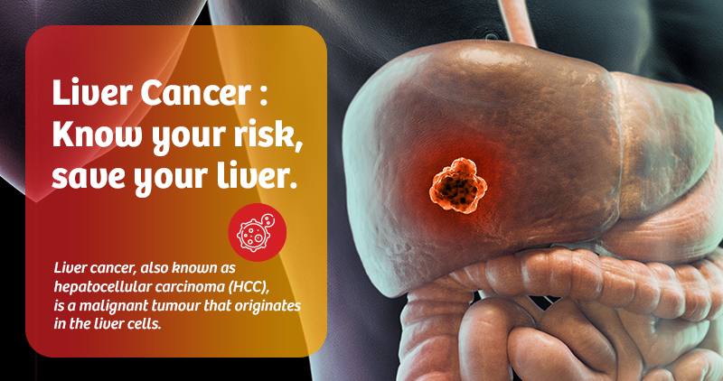
Liver cancer, also known as hepatocellular carcinoma (HCC), is a malignant tumour that originates in the liver cells. Worldwide it is the fifth most common cancer in men and ninth most common cancer in females, particularly in regions where hepatitis B and C are prevalent. This blog aims to explore liver cancer from a technical perspective, delving into its pathophysiology, risk factors, diagnostic methods, treatment options, and ongoing research developments.
Pathophysiology of liver cancer:
The liver is responsible for many vital functions, including detoxification, synthesis of proteins, and bile production. However, the liver is also susceptible to a variety of diseases that can lead to cancer. Liver cancer primarily arises from hepatocytes, the liver’s cell type.
The progression of liver cancer typically begins with liver cirrhosis or chronic liver inflammation caused by conditions like viral hepatitis (hepatitis B or C), alcoholic liver disease, and non-alcoholic fatty liver disease (NAFLD). These conditions lead to mutations and genomic instability in liver cells. Over time, these mutations accumulate, giving rise to abnormal growth and division of hepatocytes, eventually leading to the formation of a tumour.
Molecular mechanisms:
- Genetic mutations: Several genetic mutations are associated with liver cancer, including alterations in the TP53, CTNNB1, and TERT genes. These mutations drive the uncontrolled growth of liver cells.
- Epigenetic changes: Abnormal DNA methylation and histone modifications can affect gene expression without altering the DNA sequence, further contributing to tumorigenesis.
- Inflammatory cytokines: Chronic liver inflammation results in the release of cytokines like TNF-α and IL-6, which promote cell proliferation, angiogenesis (new blood vessel formation), and resistance to apoptosis (programmed cell death), all hallmarks of cancer.
Risk factors for liver cancer:
The development of liver cancer is influenced by both genetic and environmental factors. Key risk factors include:
- Chronic viral hepatitis: Chronic infection with hepatitis B virus (HBV) or hepatitis C virus (HCV) leads to liver inflammation and fibrosis, which can progress to cirrhosis and liver cancer. HBV, in particular, can integrate its genome into the host’s liver cells, directly contributing to oncogenesis.
- Alcoholic liver disease: Chronic alcohol consumption leads to oxidative stress in the liver, which leads to inflammation and cirrhosis, subsequent development of HCC. The metabolisation of alcohol produces acetaldehyde, a potent carcinogen.
- Non-alcoholic fatty liver disease (NAFLD): NAFLD is closely linked to obesity, insulin resistance, and metabolic syndrome. This condition can progress to non-alcoholic steatohepatitis (NASH), which increases the risk of HCC.
- Chemical carcinogens: Aflatoxins are toxic compounds produced by the fungus Aspergillus flavus found on improperly stored grains and legumes. These toxins cause DNA damage, leading to mutations in the TP53 tumour suppressor gene.
- Hereditary Factors: Genetic predispositions, including inherited liver diseases like hemochromatosis (iron overload) and Wilson’s disease (copper accumulation), can increase the risk of developing liver cancer.
Diagnostic approaches:
Diagnosing liver cancer involves a combination of clinical, imaging, and laboratory techniques. Early detection is challenging because liver cancer often does not present symptoms until it is in an advanced stage. Therefore, regular screening is recommended for high-risk populations.
- Imaging techniques:
- Ultrasound: The primary screening tool for liver cancer, especially in patients with cirrhosis or chronic hepatitis. It can detect liver nodules and lesions.
- Contrast-enhanced imaging CT SCAN and MRI: Characteristic findings of uptake in the arterial phase and washout in the venous phase have high sensitivity and specificity in diagnosing HCC. The American College of Radiology has proposed a system for estimating the likelihood of malignancy, in which LI-RADS 5 is malignant.
- Biopsy: A liver biopsy may be indicated for suspicious lesions when diagnosis cannot be confirmed radiologically or if alternative diagnoses are suspected.
- Serological markers:
- Alpha-fetoprotein (AFP): AFP is the most commonly used biomarker in liver cancer. Elevated levels are often seen in patients with HCC, although it is not specific and may be elevated in other conditions like cirrhosis or pregnancy. Presence of liver mass with cirrhosis and AFP > 400 ng/ml is usually indicative of HCC.
- Des-gamma-carboxyprothrombin (DCP): This is another biomarker used to diagnose liver cancer, especially in patients with chronic liver disease.
- Prothrombin induced by vitamin K absence- II (PIVKA -II) is an abnormal prothrombin protein elevated in HCC with an overall specificity of more than 80%.
Treatment options:
The treatment of liver cancer depends on various factors, including the size and location of the tumour, the liver’s overall health (Decided by Child-Pugh criteria), and whether the cancer has spread (metastasised). Treatment options can be broadly classified into:
- Surgical treatments
- Liver resection: In cases where the tumour is localised, and the liver function is still preserved, parenchyma-preserving liver resection can be performed.
- Liver transplantation: For patients with cirrhosis or multiple tumours, liver transplantation offers the best chance for survival. Transplantation is considered when the patient meets specific criteria, such as the milan criteria (a single tumour ≤5 cm or up to three tumours each ≤3 cm in the absence of portal invasion and extrahepatic disease), other extended criteria are UCSF(University of California - San Francisco)
- Ablation therapies
- Radiofrequency ablation (RFA): This technique uses an electrode inserted in a tumour and delivers a high-frequency current generating heat which destroys the tumour. It is effective in tumours <3 CM in diameter.
- Percutaneous ethanol injection (PEI): Ethanol is injected into the tumour to cause necrosis.
- Microwave ablation: Similarly, RFA uses microwave energy to destroy tumour cells.
- Interventional radiology:
- Transarterial chemoembolisation (TACE): TACE involves the selective delivery of chemotherapy drugs directly to the tumour, followed by embolisation to block the blood supply to the cancer cells.
- Transarterial radioembolisation (TARE): A more advanced version of TACE, where radioactive microspheres are used to deliver targeted radiation to the tumour.
- Radiotherapy:
- Stereotactic body radiotherapy (SBRT): it is a newer method of delivering high dose, high precision radiation in a few treatments Recently it has been investigated as a definitive treatment for smaller tumours with satisfactory results.
- Systemic therapies:
- Targeted therapy: Drugs like Sorafenib and Lenvatinib target specific molecular pathways involved in tumour growth, such as the VEGF (vascular endothelial growth factor) pathway, which is crucial for angiogenesis.
- Immunotherapy: Checkpoint inhibitors such as nivolumab and pembrolizumab are being investigated as treatment options, as they help the immune system recognise and attack cancer cells.
- Chemotherapy: Chemotherapy is not commonly used in HCC as liver cancer tends to be resistant to conventional chemotherapy agents. However, it may be considered in specific cases.
Research and future directions
The landscape of liver cancer treatment is rapidly evolving, with significant advancements in precision medicine and immunotherapy.
Ongoing research is focused on:
- Biomarker discovery: The identification of novel biomarkers for early detection, prognosis, and treatment monitoring is a major area of research. Liquid biopsy techniques are being explored to detect tumour DNA or RNA in blood samples.
- Gene therapy: Investigations into the use of gene-editing technologies like CRISPR-Cas9 aim to correct mutations in liver cells, offering a potential new avenue for treatment.
- Immunotherapy combination therapies: Combining checkpoint inhibitors with other treatments, such as targeted therapy or chemotherapy, is showing promise in enhancing the immune response against liver cancer.
Best cancer care in Gujarat.
In a nutshell:
Liver cancer remains a major global health challenge, but with continued research and advancements in molecular biology, imaging technologies, and treatment options, the outlook for patients is improving. Early detection, precise targeting of tumour cells, and personalised treatment approaches are key to improving survival rates and reducing the burden of this deadly disease. As we continue to understand the molecular mechanisms of liver cancer, we move closer to a future where better outcomes are achievable for patients worldwide.
At KD Hospital, Ahmedabad, we understand that the fight against this cancer requires a multifaceted approach. Our team of expert oncologists, surgeons, radiotherapists, and supportive care specialists collaborate to create a personalised treatment plan for your health needs. With advanced technology, surgical techniques, and compassionate patient-centred care, KD Hospital provides the best possible treatment to fight against liver cancer.

